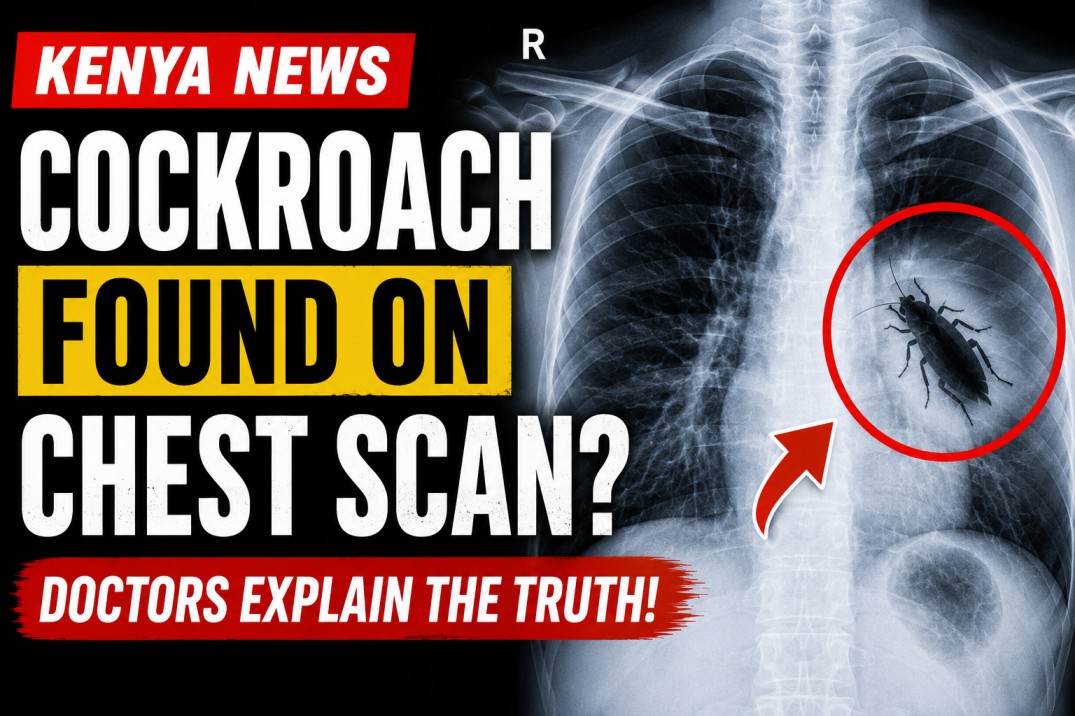
A chest scan showing what appeared to be a cockroach has gone viral after doctors in Kenya shared the unusual image online, leaving many people shocked and amused. While the scan quickly sparked speculation that an insect had somehow entered a patient’s chest, medical experts have clarified that the reality was far less alarming—and highlights the importance of careful diagnosis.
The case involved a patient who underwent a routine chest X-ray at a hospital in Kenya. The resulting image appeared to show the unmistakable outline of a cockroach over the chest area. Images of the scan circulated widely on social media, with many users expressing disbelief and curiosity.
However, healthcare professionals later explained that the cockroach was not inside the patient’s body. Instead, it had become trapped between the X-ray cassette and the detector during the imaging process, creating an artifact that appeared on the scan. Such imaging artifacts can occasionally occur when foreign objects interfere with diagnostic equipment.
Radiologists noted that X-rays produce two-dimensional images, making it possible for objects outside the body to appear as though they are inside. This is why medical imaging is always interpreted alongside a patient’s symptoms, medical history, and, when necessary, additional tests.
Experts say the incident serves as a reminder that not everything visible on a medical scan represents a disease or abnormality within the body. Dust, clothing accessories, jewelry, ECG leads, buttons, hair clips, or even insects that accidentally come into contact with imaging equipment can produce misleading shadows or shapes.
Medical professionals emphasize that quality checks are an essential part of radiology. If an unusual image appears that does not match the patient’s clinical condition, radiographers and radiologists may repeat the scan after ensuring the equipment is clean and free of external objects.
Although the “cockroach chest scan” made for an entertaining viral story, doctors say it also offers an opportunity to educate the public about how medical imaging works. X-rays are valuable diagnostic tools, but they are only one part of the diagnostic process and must always be interpreted by trained healthcare professionals.
The Kenyan case has since become an example of how unusual imaging artifacts can capture public attention while underscoring the importance of proper imaging techniques and expert interpretation. Fortunately, the patient was not found to have a cockroach inside their chest, and the startling image was simply the result of an unexpected encounter between an insect and the X-ray equipment.